Autopsy Report of Edward Garner
Other
May 19, 1975
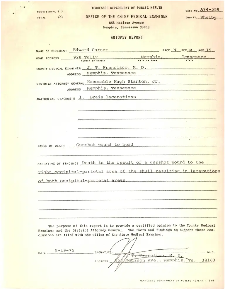
-
Case Files, Garner Working Files. Autopsy Report of Edward Garner, 1975. 512754b5-33a8-f011-bbd3-000d3a53d084. LDF Archives, Thurgood Marshall Institute. https://ldfrecollection.org/archives/archives-search/archives-item/0bd58bc3-0e75-4cf9-ad1b-0aa28c0eb592/autopsy-report-of-edward-garner. Accessed November 11, 2026.
Copied!
PROVISIONAL ( )
FINAL (X)
TEfTHESSEE OEPARTTiEHT OF PUBLIC HEALTH
OFFICE OF THE CHIEF MEDICAL EXAMINER
858 Madison Avenue
Memphis, Tennessee 33103
AUTOPSY REPORT
CASE MO A74 —5 59
couN TY__she l-tiLy—
NAME OF DECEDENT E d w a r d G a m e r
HOME ADDRESS 9 2 8 T a l l y M e m p h i s ,
RACE N SEX M AGE 1 S
_______Xen.ng.S-S.e e .
NUMSCP OK STHECT CITr OK TO»H
COUNTr’ MEDICAL EXAf.4INER J . T . F r a n c i s C O ^ __Mj:— D. _
ADDRESS M e m p h i s , T e n n e s s e e
DISTRICT ATTORNEY GENERAL H o n o r a b l e H u g h S t a n t o n , J r .
ADDRESS M e m p h i s , T e n n e s s e e
ANATOMICAL DIAGNOSIS 1 . B r a i n l a c e r a t i o n s
CAUSE OF DEATH G u n s h o t w o u n d t o h e a d
narra ti ve OF FINDINGS D e a t h i s t h e r e s u l t o f a g u n s h o t v/ound t o t h e ________
r i g h t o c c i p i t a 1 - p a r i e t a l a r e a o f t h e s k u l l r e s u l t i n . a _ . i n . l a c e r a t i o n s
o f b o t h o c c i p i t a l - p a r i e t a l a r e a s . ------ ------------- --------------------------------------------------------------------------------
The purpose o f th is report is to provide a c e r t i f ie d opinion to the County Medical
Examiner and the D istrict Attorney General. The facts and findings to support these con
clusions are fi led with the office of the State Medical Examiner.
DATE 5 - 1 9 - 7 5
T E N N E S S E E d e p a r t m e n t O r P U B L I C H E A L T H * 1 4 4
T H E C I T Y O F M E M P H I S H 0 3 P 1 T A L
A U T O P S Y P R O T O C O L
Name Edward Garner__________
service . HosDital No.
Aqe IS RaceNearn
Date of Admission Date and Hour of Death
Date and Hour of Autopsy 1 0 -4 -7 4 1 4 :0 0
Patholoqist D r s . F r a n c i s c o . B e l l Assistant Dr. S aoala
Checked by j . m. Franc i sen , M. n.________ Date Completed S -1 9 -7 S
FINAL PATHOLOGICAL DIAGNOSIS
PRIMARY SERIES:
I . Gunshot wound to r i g h t o c c i p i t o - p a r i e t a l area
A . B ra in la c e r a t i o n s
1 . R ig h t o c c i p i t o - p a r i e t a l area
2 . L e f t o c c i p i t o - p a r i e t a l area
3 . F ra c tu re d l e f t o c c i p i t o - p a r i e t a l bone
B. S t a t u s p o s t -o p e r a t i v e s u r g i c a l r e p a i r
SECONDARY SERIES;
1 . E n d o c a r d ia l p e t e c h ia e
LABORATORY FINDINGS:
Blood E t h a n o l : 0 .09%
Blood t y p e : Group O, Rh p o s i t i v e
3 6 5
P a g e 1
Autopsy
C IT Y O F MEMPHIS H O S P IT A L S
A U T O P SY P R O T O C O L
W Indicofes normal gross
Exominofion
Wf. ^ 0 lb. Ht'. Lo ̂ in
Good Poor
(T oxture)
Stofe of Nutrition
Hair (color)
Edema (degree) £)
Scars and Wounds ^
Sl<in L es io n s
Deformities
Polpable m asses
{ ) E y e s (color)
( 0-T^se
( - )^ r ;o st5
( )^£Jcfemal Genitalia and anus
Somatype
Very Poor
(Di stribution)
IN S P EC T IO N AND P A L P A T IO N
Joundice
Skin color
Special Chort Not Done
12
Cul tures
Or emi stry
( o y e r i cordium
( Pleuro
( S-Rjght Pleura
( ) Pjrntoneom
Mox. th ickness abdominal panniculus
Adhesions
Other abnormalities
Relations of viscera
( ki T̂ongue
( ^y^arotids
( TPo rto l vein
( ) Lymph nodes
( - i T h y r o i d ( w t l '- .^ O
( i)^ul monory ortery
,gm.
Light
( V ^ p i l s
( )''̂ rfk)uth
S EC TIO N
■ u
>(
sp. gr.
Thoracic-cordiac ratio
( (C&hOrynx
( "V^Lm a X i 11 o ry
{ *-V^prynx
( ' " T r o r ot hy ro i ds
( ‘■')0"hy mu s
( ophr ogm
Cyanosis
Medium Dork
Special Chart II
( L U c
character
3 6 5
( ) Heart (wt). . gfTi.
( ‘-t^picardium
( c a r d i u m
Left ventricle diloted.
( ^^^ndocardium
Pago 2
Autopsy No
Special Chart 1
Thickened
Thrombi
Right ventricle d i la te d ______
o -------------------- ------- L
th ickness.
Valve Measurement
{ ) Tricuspid________
( ) Mitral_____________
/ o th ickness.
' I
( ) Aortic. n
Fi brosi s
t K
Vegetation end Size
{ ) Pulmonic.
( •i^Caroaanes
1
Special Chart 2
(L^^ordae Tendenae.
Stenosi s Osti Lumen
degree .
Thrombi or cm boli
( Ŝ -'Aorta
Arteriosclerosis Type
Location
Lungs ( ) Right / ^ gm.
Left / ____ _ gtti.
Consolidation
Atelectasis
Degree____
Elast ic ity
Ascending
Col ci fi ed
Thorocic
Adhesion s
Location ond si ze.
Location ond s iz e _
Ulceroted
Abdominal
Speciol Chart 3
. Speci ol Cf'ort 4
3 6 7
P a g e 3
Autopsy No
O 1
( ) Spleen w t._ L C L - . 9^-
Other
( )‘-p'^phag'js Mucosa
( xLJSf^gch
/(W
Dimensions.
Ulcerated
Ulcerated
Contents. . cc .
( ) (0 y 6 6 e
( ) Recti^Fi
( txiC^svt. jooT)_
Hemorrhoi ds
gm. Dimensions ,
Thrombosed
( ) Duct stones
( ) Lobulotion
Kidney ( ) right wt._ gm.
Dimen lion 5____________ x ------------ x
C o r t e x ______________ mm.
Oth«
Special Chart 6
Special Chart 5
Special Chart 5
Speciol Chart 5
Special Chart 6
3 6 8
Pea*! ^
/ 9 7 ^ / -
Au f p iy No--------- --------
r
kidnry cont d . . .
R i g h t k i d n e y Left kidn-y
Cortical surface Smooth noduior frtol fvnoct' 1 mdijlnr fetal
F me Coarse "-int Conrse
re i VI __________________________ _________
Special Chart 7
( ) Ureters Patent Obstructed Dilat-d St-.n-s
Locotion I
(k-)^ladder Urine Suaar
Trabeciilction F l in t Moderate Prominent
( ) Prostate wf.
Obstruction 1+ 2+ 3f-
{ ) Urethra Mucosa
r j-«nr-*nObstruction
( ) Seminal v^si cle
( ) T est is f ) Right
( ) L e f t _
Specie! Chart 7
( ) Vagina
( ) Uterus
Normal
Hymen
Weight______
( ) Cervix
Atroohic
intact
Voginiti s
Ruptured
Typ"' of f lu id..
Dimensions.
( ) Endometrium T h ic k n e s s— --------— — --------------
( ) Fcllopinn T u k-s Adhorions Ohstructi-r,
Ovary ( ) Right ( ) Left Dimensions------------------- x ---------
C o r p u s L u t e u m C o r p u s A l b i c o n s Specie! Chart 7
3 ; ; 9
Page 5
Autopsy No.
Adrenals ( ) Right S ize . gm. Left Si 1 (r- . gm.
Corticol nodularity None
( ) Medulla ______________________
Other None Other.
( ) Pituitary
Infarcts
( ) Sltelotal Nl.
Lipping vertebro
Exostosi s
( ) Bono Marrow
Red
{ ) Sinuses
Si ze.
Osteoporosi s
eh ere.
Fatty
( ) Brain
( ) Sphenoid
( ) Mid. Eor
( ) Other
Not examined
.gm.
. mgm.
Tumors
Where.
Where.
C lear
W,.
( ) Dura
( ) Pia-orachnoid
( ) Venous sinuses
( ) Pineal
( ) Spinol cord w t.______________ gm.
Size Cervical C-3 mm..
Thorocic D-10 mm..
Lumbar L-3 mm..
Other______________________
Exudate
Thrombi
Special Chart 8
Purulent
Atrophy Hemorrhage
Special Chart 9
Special Churf 10
if l\.
C H A R T /(/18
/ ) 7
c r ■
^ J
RIGHT HEAD Special Chari- 14
7
̂ A 7 4 -5 5 9
Edward Garner
HEART:
LUMG :
PANCREAS:
KIDNEY:
Normal
Normal
Normal
Normal
MICROSCOPIC SUMMARY
Edward Gamer 318917 Neurosurgery
C IT Y OF MEMPHIS HOSPITALS
O' .H O S P IT A L .
Admltted-
Discharg* Summary
10-3-74
Dlschorgod. l .q- ^ - 7 4
This young black male was brought to the John Gaston Hospital
Emergency'Room the evening of 10-3-74 with a history that he had
been shot in the head by the police in circumstances which were vm-
known to me. When I first saw this young man, he was lying in the
Emergency Room with a gunshot wound on the back of the head. There
was a lot of blood on the sheet. He was moaningsome, lying on the
bed. He was bilaterally decerebrate. The pupils were midposition
and fixed. Doll's head movements appear to be intact bilaterally.
His repirations were approximately 10 per minute and quite shallow.
His pulse was about 60 and regular, the X-ray showed a gunshot
wound on the back of the head that appeared to be transcereball,
I thought on the pre-operative Xray examination there might be
a superior sagittal sinus injury in addition. Thepatient was taken
to surgery in a very critical condition. We attempted to do a
craniotomy and repair teh gunshot wound to the head; however, he
expired on teh table soon after we got surgery started. At surgery,
we did find an extensive skull injury on the back of the head, and
in addition, a transected superior sggittal sinus.
ADMISSION DIAGNOSIS: Gunshot wound to the head.
DISCHARGE DIAGNOSIS: Same, with extensive cortical injury and
superior sagittal sinus transection,
OPERATIVE AND SPECIAL PROCEDURES: Attempted repair of gunshot woundto head through craniotomy,
Howard morg^, M.D.
12-29-74
HM/DI
F O R M # 4 0 A
M A M S A M O U N I T N U M U N A O M I M I O N N U M M I I N U H O IM O O T A T IO N
DOOM on Bu> Mima
C IT Y o r M KM P H IS H O aP fT A I.
PROOREM NOTES
UNIT N O .
2 m .
/ r -
c f f T U c . ^
1 ,
73
# 7 0 0 R E V .
7/20/72 CITY OF MEMPHIS HOSPITALS
REPORT OF OPERATION
Unit No— a is a iz .
HOSPITAL Gastca.
Name Edward Garner
Date of
.Operation___10-4-74
Date of
■Dictation 10-4-74 Floor
Attending Surgeon pr. Jame s- T. Robe rtson------ Resident Surgeon — Dr. Mdrgan-
Assistants Dr. Will lams---------------------------------------- ----------------------------------------------------
Anesthetist Dr. NI&Mok-a------------------------
Anes. Time Total Anes.
Began _______________________ T im e______
Anesthetic genars--nasal tracheal-------
Scrub Circulating
— Nurse— Hayes-------------- Nurse Downey
Operation: ATTEMPTED REPAIR OF GUNSHOT WOUND TO HEAD
Sponge Count; Correct Q Missing Q Condition of patient Operating time
Preoperative diagnosis: Gunshot wound to head, tra n s p a r ie ta l , probable superior sa g it ta l
sinus in ju ry .
Postoperative diagnosis: T ran scortica l gunshot wormo tc head, p a r ie t a l , with complete
la ce ra t io n o f superior sa g it ta l sinus.
Surgical Findings: jh e e n tr 'n ce wound in the head was in the r igh t parieta l area.
There were numerous comminuted bone fragments in the p o s te r io r parietal area on both s ides
o v e r the m'.dline. The e x i t vxjund was ii. the l e f t parieta l area. The comrainutda bone
fragments were e a s i ly l i f t e d o f f o f the dura. The entrance wound in the brain was ju s t
underneath the entrance wound in the skin about two inches from the m id line . The b u l le t
traversed through the su p erior parieta l lobe and then t o t a l l y la cera ted . The superior
s a g i t t a l s inus. There was considerab le amount o f c o r t i c a l damage done oi. the l e f t s ide
in the parieta l lobe a ls o .
PROCEDURE: This young black lale was in a very c r i t i c a l s ta te having recently been shot
in the head. He had been f i r s t seen in the Emergency Room where h is neurolog ica l status
was grave. He vas given Mannitol, x-rays taken o f the skull and he was immediately
rushed to surgery. He was i i tu b r te d nasal t ra ch e a lly end placed on the operating tab le
in the prone p o s i t io n , with the head resting on the ce r e b e l la r head r e s t . P os te r io r aspect
o f the head was shaved, prep 'd and draped in the s-^andard fash ion . The entrance wound
as mentioned was in the r ig h t parieta l area with the e x i t wound in the l e f t p ar ie ta l area.
An in c is io n was made in the coronal plane incorporating these two la cera t ion s where the
b u l l e t had penetrated. The skin f la p was then flapped back s u b p e r io s te a l ly . As mentioned
we encountered a v;hole bunch o f comminuted fragments o f s k u l l . Thes'. were l i f t e d o f f
the dura and we could see immediately that tha b u l le t had traversed the superior s a g it ta l
s in u s and had t o t a l l y la cerated th is s tru ctu re . Bleeding v/as vigorous f o r ju s t a few
m inutes, but i t was e a s i ly c o n tr o l le d with f in g e r pressure on the s inus. At th is time the
p a t ie n t who had not had very good v i ta l signs at th is point began to become worse and
he suddenly was without blood pressure and pulse. At th is point we l i g a t 'd the
s u p e r io r sa g it ta l sinus and c lo se d the wound rap id ly with s i l k and nylon. Estimated blood
l o s s 'uring the surgical procedure was about one un it . This amount o f blood v.as replaced
p r i o r to his demise. The patien t was taken to the intensive care unit where he wi l l be
s e n t to the morgue soon.
Howard Morgan, M. D.Pdy